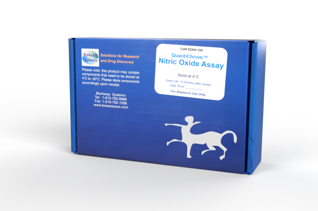
DESCRIPTION
Nitric oxide (NO) is a reactive radical that plays an important role in many key physiological functions. NO, an oxidation product of arginine by nitric oxide synthase, is involved in host defense and development, activation of regulatory proteins and direct covalent interaction with functional biomolecules. Simple, direct and automation-ready procedures for measuring NO are becoming popular in Research and Drug Discovery. Since NO is oxidized to nitrite and nitrate, it is common practice to quantitate total NO2 - /NO3 - as a measure for NO level. BioAssay Systems' QuantiChromTM Nitric Oxide Assay Kit is designed to accurately measure NO production following reduction of nitrate to nitrite using improved Griess method. The procedure is simple and the time required for sample pretreatment and assay is reduced to as short as 30 min.
KEY FEATURES
Sensitive and accurate. Detection range 0.6 - 100 µM in 96-well plate. Rapid and reliable. Using an optimized VCl3 reagent, the time required for reduction of NO3 - to NO2 - is 10 min at 60°C. Simple and high-throughput. The procedure involves mixing sample with three reagents, incubation for 10 min at 60°C and reading the optical density. Can be readily automated to measure thousands of samples per day.
APPLICATIONS
Direct Assays: NO in plasma, serum, urine, tissue/cells and foods. Drug Discovery/Pharmacology: effects of drugs on NO metabolism.
KIT CONTENTS (100 tests in 96-well plates)
Reagent A: 12 mL Reagent B: 500 µL Reagent C: 12 mL
NaOH: 1 mL ZnSO4: 1 mL Standard: 1 mL
Storage conditions. The kit is shipped at room temperature. Store the Standard at -20°C and all other reagents at -20 to 4°C. Shelf life of six months after receipt.
Precautions: reagents are for research use only. Please refer to Material Safety Data Sheet for detailed information.
PROCEDURES
Prior to assay, equilibrate all components to room temperature. If
precipitates are present in Reagent B, warm at 37˚C until redissolved
(~10-15 min).
Sample treatment: tissue or cell samples are homogenized in 1 x PBS (pH 7.4). Centrifuge at 14,000 rpm at 4°C. Use supernatant for NO assay.
Samples that need deproteination include serum, plasma, whole blood, cell culture media containing FBS, tissue or cell lysates. Urine and saliva do not need deproteination.
Deproteination. Mix 150 µL sample with 8 µL ZnSO4 in 1.5-mL tubes. Vortex and then add 8 µL NaOH, votex again and centrifuge 10 min at 14,000 rpm. Transfer 100 µL of the clear supernatant to a clean tube. Note: If samples need to be deproteinated, 150 µL of each standard should be prepared and also treated with ZnSO4 and NaOH to eliminate the need for a dilution factor.
Procedure using 96-well plate:
1. Standards. Prepare 500 µL 100 µM Premix by mixing 50 µL 1.0 mM Standard and 450 µL distilled water. Dilute standards in 1.5 mL centrifuge tubes as described in the Table.
2. Reaction. Add 100 µL of Standard and Sample to separate, labeled eppendorf tubes. (We recommend that samples be measured in at least duplicate). Immediately prior to starting the reaction, prepare enough Working Reagent (WR) for all samples and standards by mixing per reaction tube: 100 µL Reagent A, 4 µL Reagent B and 100 µL Reagent C. Add 200 µL of the WR to each sample and standard tube and incubate for 10 min at 60°C. (Alternatively, the reaction can be run at 37°C for 60 min or RT for 150 min.) Note: If precipitates are present in Reagent B, warm at 37˚C until redissolved (~10-15 min).
3. Measurement. Briefly centrifuge the reaction tubes to pellet any condensation and transfer 250 µL of each reaction to separate wells in a 96 well plate. Read OD at 500-570 nm (peak 540 nm).
Procedure using Cuvette:
Prepare standards and samples as described for the 96-well procedure except quadruple (4x) the volumes. After the reaction, transfer 1 mL to a cuvette. Measure OD540nm in the cuvette.
CALCULATION
Subtract blank OD (Std 4) from the standard OD values and plot the OD against standard concentrations. Determine the slope using linear regression fitting. The NO concentration of Sample is calculated from the Slope of the standard curve as
ODSAMPLE and ODBLANK are optical density values of the sample and water, respectively. If calculated nitric oxide concentration is higher than 100 µM, dilute sample in water, repeat assay and multiply the results by the dilution factor n.
Conversions: 1 mg/dL NO equals 333 µM, 0.001% or 10 ppm.
MATERIALS REQUIRED, BUT NOT PROVIDED
Pipetting devices, eppendorf tubes, eppendorf centrifuge, clear, flat bottomed 96 well plates or cuvettes, plate reader or spectrophotometer and heat block or hot water bath (optional).
GENERAL CONSIDERATIONS
Antioxidants and nucleophiles (e.g. β-mercaptoethanol, glutathione, dithiothreitol and cysteine) may interfere with this assay. Avoid using these compounds during sample preparation.
PUBLICATIONS
1. Kim, H.-C., et al. (2019). Cobalt(Ii)-coordination polymers containing glutarates and bipyridyl ligands and their antifungal potential. Scientific Reports 9(1).
2. Kang, M., et al. (2020). Plasma mediated disinfection of rice seeds in water and air. Journal of Physics D: Applied Physics 53(21).
3. Veerana, M., et al. (2021). Analysis of the effects of Cu-MOFs on
fungal cell inactivation. RSC Advances 11(2): 1057-1065.
