DESCRIPTION
CREATININE is synthesized in the body from creatine, which is produced during muscle contractions from creatine phosphate. In the blood, creatinine is removed by filtration through the glomeruli of the kidney and is secreted into urine. In healthy individuals, creatinine secretion is independent of diet and is fairly constant. The creatinine clearance test has become one of the most sensitive tests for measuring glomerular filtration rate. In kidney disease, creatinine levels in the blood are elevated, whereas the creatinine clearance rate and hence the urine levels are diminished. Creatinine testing is most widely used to assess kidney function.
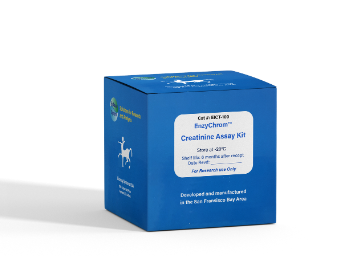
BioAssay Systems’ enzyme-based creatinine assay uses a reaction sequence that excludes endogenous ammonia to convert a dye into a colored and fluorescent form. The absorbance at 570 nm or fluorescence intensity at λex/em = 530/585 nm is directly proportional to the creatinine concentration in the sample.
KEY FEATURES
Fast and sensitive. Linear detection range: 4.8 to 500 µM or 0.054-5.7 mg/dL (colorimetric assay) and 0.25 to 100 µM or 0.0028-1.14 mg/dL (fluorimetric assay) for a 60 min reaction.
Convenient. The procedure involves adding a single working reagent and reading after 60 minutes. Room temperature assay. No 37ºC heater is needed.
High-throughput. Homogeneous "mix-incubate-measure" type assay. Can be readily automated to process thousands of samples per day.
APPLICATIONS
Creatinine determination in urine, serum, plasma, and other biological preparations.
KIT CONTENTS (100 TESTS IN 96-WELL PLATES)
Assay Buffer: 20 mL Enzyme A: Dried
Enzyme B: 120 µL Dye Reagent: 120 µL
Standard: 1 mL
Storage conditions. The kit is shipped on ice. Store all components at -20°C upon receiving. Shelf life: 6 months after receipt.
Precautions: reagents are for research use only. Normal precautions for laboratory reagents should be exercised while using the reagents. Please refer to Safety Data Sheet for detailed information.
PROCEDURES
Sample Preparation. This assay is based on creatinase and creatininase reactions, creatine in the sample can interfere with the assay. If desired, creatine concentraton can be determined using our creatine assay kit (cat# ECRT-100). Urine samples should be diluted at least 100-fold with dH2O before the assay. Serum and plasma samples should be deproteinated by centrifugation for 15 min at 14000 rpm at room temperature through a 10 kDa spin filter (e.g. Corning Cat# 431483). The filtrate can be assayed directly.
Reagent Preparation. Equilibrate all reagents to room temperature before assay. Briefly centrifuge tubes before opening.
Colorimetric Procedure
1. Standards. Prepare 200 µL of 500 µM Premix by mixing 50 µL of the Standard (2 mM) and 150 µL dH2O. Dilute standards in 1.5-mL centrifuge tubes as described in the Table. Transfer 20 µL Standards into separate wells of a clear, flat bottom 96-well plate.
2. Reconstitute Enzyme A with 120 µL Assay Buffer. Pipette until all dried enzyme is dissolved. Prepare enough Working Reagent (WR) for all assay wells by mixing, for each well, 82 µL Assay Buffer, 1 µL Enzyme A, 1 µL Enzyme B, and 1 µL Dye Reagent.
Note: Reconstituted Enzyme A is stable for at least 1 month when stored at -20ºC. Add 80 µL of the WR to each well. Tap plate briefly to mix.
3. Incubate at room temperature for 60 min. Use a plate reader to read OD570nm.
Fluorimetric Procedure
1. Dilute the standards prepared in Colorimetric Procedure 1:5 in dH2O. Transfer 20 µL standards and 20 µL samples into separate wells of a black 96-well plate.
2. Add 80 µL of the WR to each well (see Colorimetric Procedure step 2). Tap plate to mix.
3. Incubate protected from light for 60 min at RT and read fluorescence at λex/em = 530/585 nm.
CALCULATION
Subtract blank value (water, standard #4) from the standard values and plot the adjusted values against standard concentrations. Determine the slope and calculate the creatinine concentration of Sample as follows
where RSAMPLE and RBLANK are the OD or fluorescence values of the Sample and H2O Blank, respectively. n is the sample dilution factor.
Conversions: 1 µM creatinine equals 0.0113 mg/dL.
MATERIALS REQUIRED, BUT NOT PROVIDED
Pipetting devices and accessories (e.g. multi-channel pipettor, pipette tips, etc), clear (colorimetric) or black (fluorimetric) flat-bottom 96-well plates, centrifuge tubes, and plate reader.
LITERATURE
1. Perrone, R.D., et al. (1992) Serum creatinine as an index of renal function: new insights into old concepts. Clin Chem, 38:1933-1953.
2. Tietz, N.W. (1999) Fundamentals of Clinical Chemistry, 3rd Ed. W. B. Saunders Company, pp. 1204-1270.
3. Kasiske, B.L., Keane, W.F. (2000) Laboratory assessment of renal disease: clearance, urinalysis, and renal biopsy. The Kidney, 6th Ed., WB Saunders Company, pp. 1129-1170.
Related Products:
QuantiChrom™ Creatinine Assay Kit (DICT-500).
